Load more
On Sale
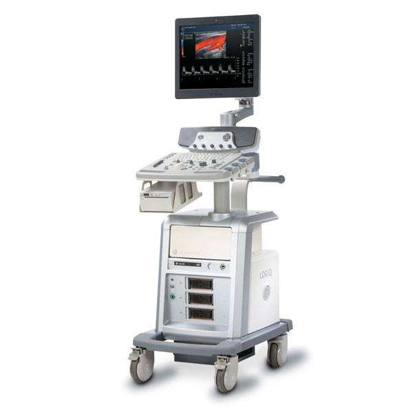
USED GE LOGIQ P6


One probe included
Main Features
- Hemodynamics is seen with great detail due to the B-Flow Color which allows you to recognize the different flow variations while maintaining high frame rates.
- Remarkable temporal and spatial resolution when scanning vessels to the imaging blood reflectors.
- Dynamic, portable, and excellent images.
- Harmonics increase the resolution and cystic clarity that incorporates coded harmonics with Phase Inversion Harmonics.
- Tissues and border differentiation are improved by the CrossXBeam. In addition, the real-time spatial compounds the acquisition and processing.
- The Raw Data promptly stores the data of the image chain to allow the best flexibility during post-processing and analysis.
- High frames rates are maintained by the DualBeam despite using high line density, even in abnormal settings. Temporal resolution is elevated in the fast-flow cardiac and vascular studies.
- The clarity of the organs and lesions are magnified by the Speckle Reduction Imaging (SRI). The high-definition contrast resolution suppresses speckle artifact while preserving true tissue architecture.
- No matter the size or difficulty of the patient every exam receives a clear and precise image. The new ceramic materials in the Single-crystal transducers expands the bandwidth and enhances the sensitivity to deliver excellent cardiac performance.
- Real-time 4D imaging achieves and produces volumetric images that envisions the multi-planar anatomical views.
System Details
- Depth: 640 mm
- Monitor: 17-inch TFT LCD
- Height: 1360 mm
- Weight: approx. 80 kg (176 lb.)
- Width: 430 mm
Operations
- Gynecological
- Vascular
- Breast
- Endocavity
- Transesophageal
- Obstetrical
- Cardiac
- Small Parts and Superficial
- Pediatric and Neonatal
- Abdominal
- Intraoperative
- Transcranial
- Urological
- Musculoskeletal
Transducer Types
- Linear Array
- Volume ‘4D’
- Convex Array
- TEE Multiplane Sector Array
- Sector Phased Array
- Single CW (Pencil)
- Bi-plane Microconvex Array
- Microconvex Array
Operating Modes
- M-Mode
- PW Doppler w/High PRF
- Anatomical M-Color Mode (option)
- Coded Contrast Imaging (option)
- Tissue Velocity Imaging (TVI) Mode (option)
- 4D Real time (option)
- Coded Harmonic Imaging
- Power Doppler Imaging (PDI) with Directional Map
- Anatomical M-Mode (option)
- B-Flow Color Mode (option)
- PFD Mode (option)
- 3D Static (option)
- Elastography (option)
- B-Mode
- Color Flow Mode (CFM)
- M-Color Flow Mode
- B-Flow Mode (option)
- CW Doppler Mode (option)
- 3D/4D Volume Modes (option)
Optional Transducers
- GE 8C – 4-11MHz, Microconvex
- GE 3CRF – 2-4MHz, Microconvex
- GE 9L – 3-10MHz, Linear
- GE T739 – 4-12MHz, Linear (Intraoperative)
- GE 3Sp – 1.5-5.5MHz, Phased Array Sector
- GE 5S – 2-5MHz, Phased Array Sector
- GE ERB – 3-11MHz, Bi-plane Intracavity
- GE 4DE7C – 4-11MHz, Convex Volume
- GE P6D – 5.0MHz, Non-Imaging Single CW Doppler Pencil
- GE 5CS – 2-6MHz, Convex
- GE E8C – 4-11MHz, Microconvex (Endocavity)
- GE BE9CS – 4-11MHz, Bi-plane Microconvex
- GE 11L – 5-13MHz, Linear
- GE 8L – 4-11MHz, Linear
- GE P2D – 2.0MHz, Non-Imaging Single CW Doppler Pencil
- GE 4C – 1.4-5.0MHz, Convex
- GE 4D8C – 4-11MHz, Microconvex Volume
- GE 4D5C-L – 3-7MHz, Convex Volume
- GE E8CS – 4-11MHz, Microconvex
- GE 4D3C-L – 2-5MHz, Convex Volume
- GR 6Tc – 3-7MHz, Multiplane Transesophageal Phased Array Sector
- GE 3S – 1.5-3.5MHz, Phased Array Sector
- GE 7S – 3-8MHz, Phased Array Sector
- GE 5Sp – 4-10MHz, Phased Array Sector
- GE i12L – 5-12MHz, Linear (Intraoperative)
- GE i739 – 4-12MHz, Linear (Intraoperative)
- GE ML6-15 – 6-15MHz, Linear