Load more


On Sale
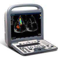
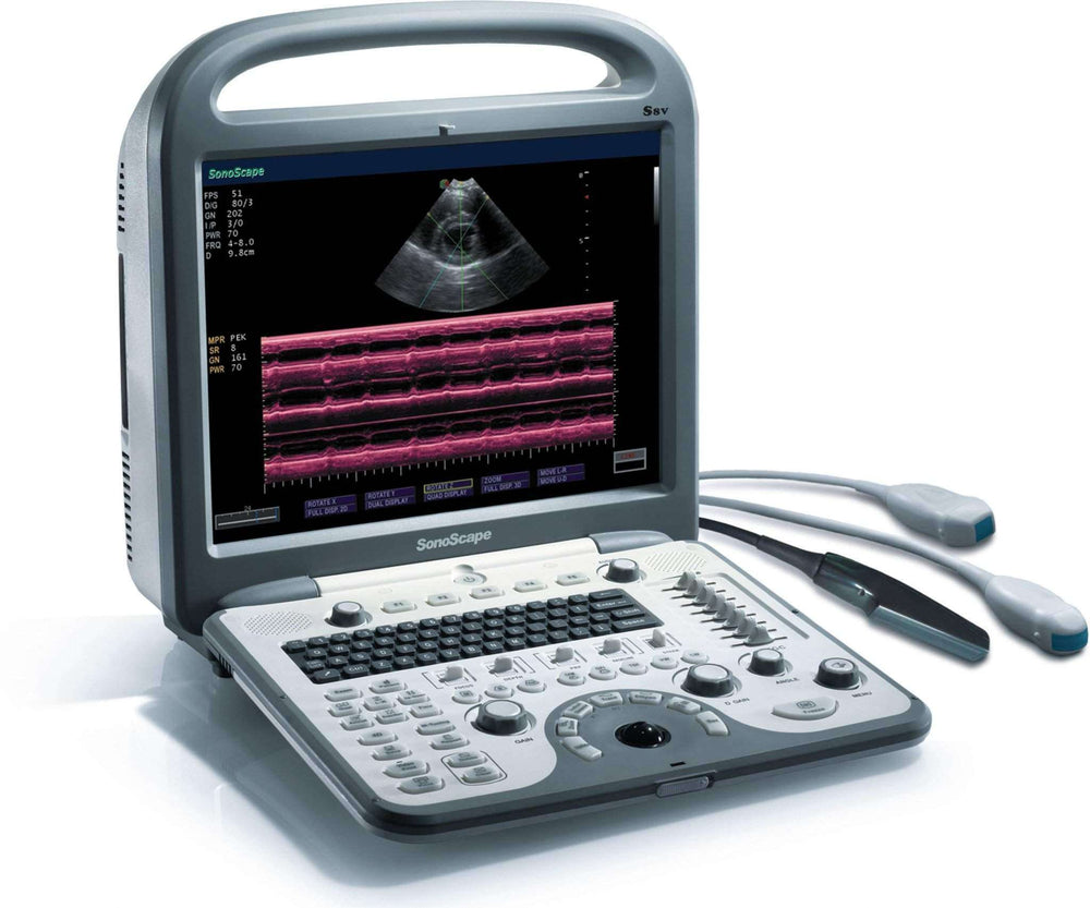
SonoScape S8 Demo Model


Comes with one probe - Deal!
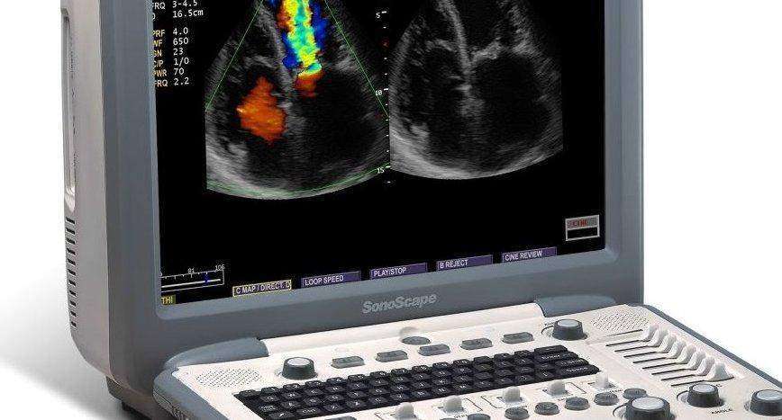
We used this model for an hour or so to take pictures. Like brand new! Full Warranty.
S8 is a high-end color Doppler HCU (Hand-Carried Ultrasound) from SonoScape. This system clearly expresses the words of "an ever-higher standard of Echo HCU'. Although S8 is a HCU system, its performance is equal to high-end cart-based echo ultrasound systems. This flagship HCU is incorporated with high configuration to meet the strictest requirements from cardiologist, such as adult TEE, pediatric TEE, high frequency phased array, 15 MHz linear transducer, 512 super high density transducers, 4D imaging function, and so on.
Features
- HCU design with two transducer sockets; Convertible trolley design
- 15" LCD monitor with super large imaging area
- Full cardiovascular transducers: TEE, Pediatric TEE, Phased array, High frequency phased array, High-frequency linear, etc.
- High density transducers with frequency ranges from 1.9 to 15MHz
- Integrated with state-of-the-art technologies, like μ-scan technology, IMT, multiple-beam processing, automatic flow volume analysis, 4D imaging technology, etc.
- Comprehensive cardiovascular analysis kits: TDI, Steer M, Color M; CW, HPRF, Panoramic image, etc.
- Full patient database solutions: DVD RW, DICOM3.0, AVI/JPG, USB2.0, HDD, PDF report, etc.
- Built-in high capacity Li-ion battery
- Liver. The ultrasonic examination of liver is a relatively high informative method. The doctor evaluates the dimensions of the liver, its structure and homogeneity, local disturbances as well as the blood flow condition. The ultrasonic examination allows to detect both diffusive changes in the liver (liver steatosis, chronic active liver disease, cirrhosis) and local (fluid formalities and tumor mass).
- The gall bladder and bile passages. Besides the liver itself, the state of gall bladder and bile passages is also examined: their dimensions, the thickness of the paries, patency, the presence of concrements, the state of the connective tissue. In most cases the ultrasonic examination allows to detect the concrements in the gall.
- Pancreatic gland. During the examination of the pancreatic gland the doctors can evaluate its dimensions, form, boundaries, homogeneity of parenchyma, the presence of formalities.
- Kidney and atrabiliary capsules, retroperitoneal space. During the examination of the kidney the doctors evaluate their number, arrangement, dimensions, form, boundaries, structure of parenchyma and pelvicalyceal system. The ultrasonic examination allows to detect the anomalies of the kidney, the presence of concrements, fluid formalities and tumor mass as well as changes resulting from inveterate and pathological lancinating processes in kidney. The ultrasonic examination can be used for detection of the indication of intestinal obstruction and indirect indication of adhesive process. With the help of ultrasonic examination the doctor can detect the presence of free liquid in the abdominal cavity (if it is in a great number) that can play an important role in the treatment policy of a number of therapeutic and surgical disorders and injuries.
- Thyroid gland. The ultrasonic examination is the crucial in the examination of the thyroid gland because it allows to detect the presence of ganglions, hydatids, the changes in the dimensions and structure.
- Cardiology, cardiosurgery. Echocardiography is the ultrasonic diagnostics of the heart diseases. This examination evaluates the dimensions of the heart and its particular structures (ventricle of the heart, auricle, ventricular septum, thickness of ventricular myocardium, auricle etc.) the presence and the volume of fluid in the pericard – “pericardial sac”, the state of the valves. With the help of special calculations and sizing echocardiology allows to detect the heart mass, contractility of the heart - ejection fraction, etc. There are special guides that help to observe during the operation the state of the mitral valve situated between the ventricle and auricle.
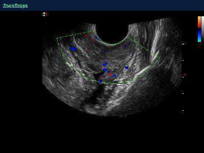
- Tocology, gynecology, prenatal diagnostics. The ultrasonic examination is used for examination of the internal female genital neoplasms, the state of the gravid uterus, anatomy and monitoring of the Embryo-fetal development. This effect is widely used in the tocology because the sound from the venter can be easily registered. At early stages of pregnancy the sound gets through the bladder. When the venter is filled with the fluid it starts to conduct the sound. The position of the placenta is detected by the sounds of blood getting through it, and within 9-10 weeks from the moment of the fetation the doctor can listen to the beating of its heart. The number of foetuses and the death of the foetus can be detected with the help of ultrasound examination.
- Muscle and skeletal examinations. The ultrasonic examination is used to detect traumatic injuries and inflammatory disease of joints (shoulder, knee, etc.), muscles, bands, meniscus, chorda, arthrosis.